Technology / Solution
Read Beyond
the Visible
Compact optics born from bio-inspired research have evolved into cameras, software and spectral vision.
01 / BIO-INSPIRED ORIGIN
Vision technology
born from bio-inspired research
Micropix’s technology began with bio-inspired lens and camera research at the KAIST Biophotonics Laboratory. Drawing on insect compound eyes, the research explored new ways of capturing images through arrays of small lenses.
The visual structure of Xenos peckii inspired ultrathin camera research using small optical channels.

02 / MLA CAMERA PLATFORM
Small lenses,
one camera
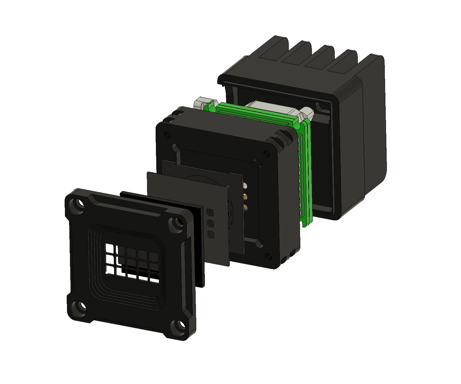
Building on this research, Micropix develops MLA-based cameras and image processing software. We connect optical design, hardware and image processing to implement the vision solutions our customers’ systems need.
03 / SPECTRAL EXPANSION
Beyond shape and color,
into the spectrum
Spectral vision is one direction for expanding the MLA camera platform. By combining spectral filters with an MLA, we capture wavelength-specific images and analyze spectral differences to reveal clues about composition and condition that RGB images alone may not distinguish.

Capture multiple spectral bands in a single shot.
Visualize captured data and analyze the target’s spectral characteristics.
04 / FROM SPECTRUM TO CLASSIFICATION
Turning spectral differences
into evidence for classification
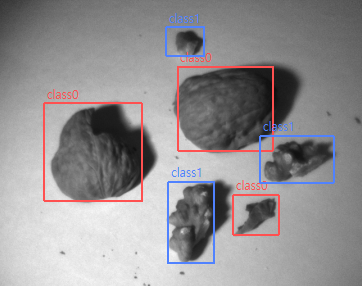
Walnut shells and kernels that appear the same color in RGB images can have different spectra. By analyzing these spectral differences and training on the data, we classify shells and kernels in the image.
RGB image
Walnut shells and kernels that appear the same color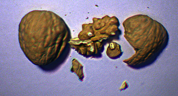
Spectral analysis
Analysis of spectral differences between two materials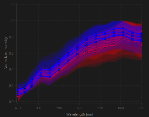
Classification
Shell and kernel classification after training